Thanks to advances in neuroscience a new research field is emerging, one which studies spiritual phenomena through neural images. It could be argued that science and technology, as understood currently in the mainstream, might not be suitable for investigating the spiritual realm. This is because different paradigms of enquiry apply. However, interdisciplinarity is leading to a blurring of boundaries and increasing numbers of science scholars are practicing meditation, while a growing number of meditators are studying and using the latest neurotechnology.
Scientific inquiry and spiritual experiences both offer a path to discovering truth. Here we offer an introduction to the study of spiritual practices as understood through the observation of neural imaging of live patients. The study of psychology and cognition is becoming increasingly relevant to our work in understanding and designing sophisticated, intelligent systems.
Most individuals, irrespective of background, nationality, culture, or age are, at some point in life, confronted with the notion of spirituality. This is often not necessarily associated with a religion or a philosophy, but rather just to find emotional wellness and balance, or to cope with life’s stresses and tribulations.
Spiritual experiences are generally associated with the heart—not the physical organ, but what the wisdom traditions consider to be our emotional center (heart chakra), which science does not study. There is nothing in the heart as a physical organ that displays association with emotions, because from a physical viewpoint, the heart is merely an involuntary muscle that pumps blood around the body—a mechanical device with a mechanical function. What makes the heart the sense organ for love and sorrow, joy and pain? Some evidence suggests that negative emotions may influence the development of chronic heart disease.*
The brain is usually considered to be the physical seat of the mind. Thanks to advances in neuroimaging, observations of the brain through sophisticated new techniques offer extremely exciting new insights. Nonetheless, the brain was an object of study long before modern technologies became widely available to make direct observations.**
The most common technologies used today to map brain and cognitive functions and mental states are based on either nuclear medicine using radioactive tracers (radiopharmaceuticals)—e.g. single photon emission computed tomography (SPECT) or positron emission tomography (PET)—and, more commonly, magnetic resonance imaging (MRI) for scanning the anatomical structure of the brain and FMRI (functional magnetic resonance) for imaging metabolic functions.
Which spiritual practices?
There is no discrete way to distinguish what is often loosely described as a spiritual practice from other areas of human mental activities that can induce certain brain states—for example, listening to music.
Scientists have tried to come up with classification systems for meditation, including a recent taxonomy. However, each method has its own limitations. Research suggests that distinct patterns of brain activation and deactivation can be identified in at least four common styles of meditation (focused attention, mantra recitation, open monitoring, and compassion/loving-kindness). Furthermore, some differences can be found among three others (visualization, sense-withdrawal, and non-dual awareness practices), showing that some brain areas are recruited consistently across multiple techniques—including insula, pre/supplementary motor cortices, the dorsal anterior cingulate cortex, and the frontopolar cortex—but convergence is the exception rather than the rule.***
Brain mapping
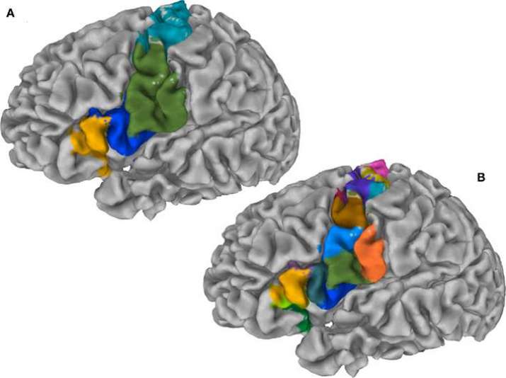
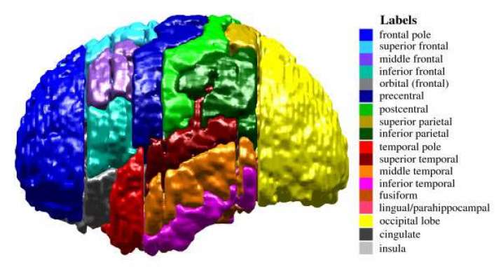
In vivo brain mapping is becoming more common thanks to the diffusion of data and relatively accessible technologies that enable observations of live patients, however the practice of studying the brain by segmenting it into regions, known as parcellation, has existed for more than a century, although it was carried out generally ex vivo (on dead patients).
The earliest brain map, which has served as the basis for neuroscience to the present day, is attributed to Korbinian Brodmann,who identified 52 regions of the brain. The earliest examples of in vivo observations are attributed to Angelo Mosso, a physiologist who monitored pulsations of the brain in adults through surgically created holes in the skull, observing that when the subjects engaged in tasks such as mathematical calculations the pulsations of the brain increased, establishing a correlation between blood flow and brain function. Currently, there are many models of brain parcellation in use and some examples can be found in Bohland et al.

What can be observed?
There are different levels of observation. Initially, studies of the brain revealed that certain areas of the brain become more “active”—where activity is defined both as chemical and electrical variations of intensity, during certain meditative practices. For example, neuro-imaging studies show that meditation results in an activation of the prefrontal cortex, activation of the thalamus and the inhibitory thalamic reticular nucleus and a resultant functional deafferentation of the parietal lobe. The other level is chemical, where different neuroimaging techniques identify chemical variations, behaviors, and changes of state generally associated with blood circulation, heat, activation, and deactivation of certain types of cell patterns and brain regions. In turn, these are associated with different cognitive events.
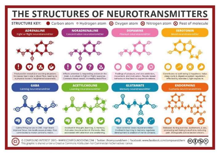
Meditative practices affect the whole brain and can be detected as neurochemical changes in the neurotransmitter network, leading to the conclusion that meditative practices cause neurophysiological and neurochemical alterations. In particular, certain mental activities typically associated with meditation and spiritual practices can result in variations of some chemicals in the brain as in the case of certain neurotransmitters—molecules associated with synapses. Changes in levels of these chemicals can trigger different behaviors (e.g. depolarization or hyperpolarization) and these chemicals can act as a neurotransmitter or a neuromodulator. There are a number of major known neurotransmitters described in the chart below.

Notably, research points to meditative practice increasing levels of dopamine and serotonin, which when low can cause depression and anxiety. Increased serotonin can also interact with dopamine, enhancing feelings of euphoria that are experienced during meditation. Serotonin in conjunction with glutamate can result in the release of acetylcholine, which is associated with attention and spatial orientation. In different studies, meditation has also been associated with changes in levels of noradrenaline and amino acid neurotransmitters, such as glutamate and GABA. One could also mention the association of meditation with an increase in plasma melatonin and various effects on the pineal gland. The bottom line is that mindfulness, associated with all meditation and spiritual practices, by inducing certain chemical changes, may have an impact on human evolution by shaping the neocortex, a phenomenon known as neuroplasticity. ****
The studies
Despite a vast and growing body of research, there is no consistent conceptual structure or method to identify the boundaries between different spiritual practices. While we do not need neuroscience to figure out that certain states, such as mental anxiety, can impact blood pressure and coronary health, research has shown that spiritual practices are not merely placebos and can cause measurable physical effects. With this knowledge, when meditators sit down to practice, they can do so with a new level of awareness of what is happening to their brain, and maybe one day they will even see the physical effects of their meditation on a screen in real time.
Science and spirituality can perhaps be associated with at least some data to back it up. The main changes for future development is to establish robust and valid research protocols and adequate methods to cope with vast amounts of data and multiple viewpoints. Eventually, if we are good enough, we might be able to demonstrate a correlation between mindful practices and human evolution.
* Kubzansky Laura D and Ichiro Kawachi. 2000. “Going to the heart of the matter: do negative emotions cause coronary heart disease?” Journal of Psychosomatic Research. Volume 48, Issues 4–5, pages 323-337
** Raichle, Marcus E. 2008. “A brief history of human brain mapping” Review Historical Perspective. Volume 32, Issue 2, pages 118–26.
*** Fox, Kieran C.R., et al, 2016. “Functional neuroanatomy of meditation: A review and meta-analysis of 78 functional neuroimaging investigations” Neuroscience & Biobehavioral Reviews. Volume 65, June 2016, Pages 208–28.
**** Davidson, Richard J, and Antoine Lutz. 2008. “Buddha’s Brain: Neuropasticity and Meditation.” IEEE Signal Process Mag. 25(1), pages 176–84.
References
Bohland, Jason W. et al. 2009. “The Brain Atlas Concordance Problem: Quantitative Comparison of Anatomical Parcellations” PlosOne
Mohandas E. 2008. “Neurobiology of Spirituality.” Mens Sana Monogr. 2008 Jan–Dec; 6(1): 63–80.
See more
FMRI Functional Magnetic Resonance Imaging Lab (California State University Long Beach)


